Module 2: Lab 2 Introduction to Microscopy using Eukaryotic Organisms
Section outline
-
 Microscopy is the scientific technique to observe objects that are too small to be seen by the naked eye. Microscopes are the tools that magnify (the process of enlarging the appearance of an object) and resolve (distinguishing two points as being separate) the minute structures such as cells, bacteria, protozoa, and their contents.
An object must measure about 100 micrometers (µm) to be visible to an average human eye without a microscope. Typical animal cells measure roughly 10 µm across but are still microscopic (require a microscope to be visible). Protists, while diverse in size, generally range from 5 micrometers to 2-3 millimeters in length, Bacterial cells are typically about 1 µm, and viruses can be 10 times smaller than bacteria.
In this course, you will learn the skill of using light microscopy to identify microorganisms (too minute to see by the naked eye but visible through microscopy). You may be required to prepare wet mount slides using an environmental sample such as cultured pond water to identify eukaryotic microorganisms. Your institution may present you with prepared dry-mount slides to study the pathogenic (disease-causing) microorganisms in humans.
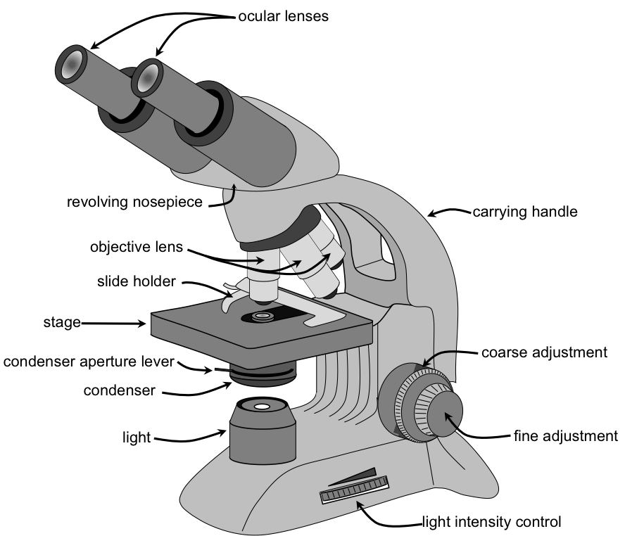
Upon completion of this module, you will be able to:- Identify the parts of a compound light microscope and describe their functions. (Course Objective# 1)
- Demonstrate correct use of the microscope, including focusing, adjusting lighting, and using oil immersion. (CLO#1)
- Prepare and observe wet mounts and stained slides of eukaryotic cells (e.g., protozoa, fungi, human cells).(CO#2,3)
- Differentiate between prokaryotic and eukaryotic cells based on structural features visible under the microscope.(CO#1, 4)
- Recognize common eukaryotic microorganisms, such as Candida albicans (fungus) or Amoeba and Paramecium (protozoa). (CO#4)
- Apply proper lab safety and cleaning protocols when handling biological samples and microscopes. (CO#1,3,4,5)
- Relate microscopic observations to clinical relevance in infection, immunity, and patient care. (CO#1,6)
To achieve these objectives: [Edit these items to match your resources and activities.]
- Read the Module 2 Introduction
- Read Chapter # in Textbook Title, Article Title, etc. [Include all reading assignments here that are outside of Moodle. Be as concise as possible. More information can be included in the third-party section below, if necessary.]
- Complete the [specific activities in the module. Include all in the order you want them completed. If an activity supports the development of foundational and/or industry-specific digital skills, note the skill to which it aligns]
Module Pressbooks Resources and Activities
You will find the following resources and activities in this module at the Pressbooks website. Click on the links below to access or complete each item.
Background Colour
Font Face
Font Kerning
Font Size
Image Visibility
Letter Spacing
Line Height
Link Highlight
Text Colour