Module 8: Urinary
Section outline
-
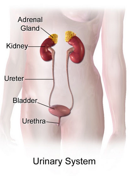
Image credit: A medical illustration depicting urinary system. August 2017. BruceBlus cc-Share Alike-4.0
Welcome to the study of the urinary system, a vital regulatory network that works continuously to maintain the body’s internal balance. Far more than a waste-removal system, the kidneys precisely regulate blood composition by controlling fluid levels, electrolytes, acid–base balance, and blood pressure, while also supporting red blood cell production and Vitamin D activation.
Introduction
The urinary system is essential for preserving the body’s internal stability by constantly monitoring and adjusting the chemical makeup of the blood. Although it is often thought of primarily as the system responsible for producing and excreting urine, its role is much broader. The kidneys regulate water balance, electrolyte levels, acid–base status, and blood pressure, and they also support red blood cell formation and the activation of Vitamin D. Collectively, these functions are fundamental to maintaining homeostasis and ensuring the proper operation of all other organ systems.
These regulatory processes occur at the microscopic level within millions of tiny functional units known as nephrons. Each nephron begins with the glomerulus, a network of capillaries that filters the blood largely according to particle size, preventing cells and most proteins from passing into the filtrate. As the filtrate moves through the nephron’s specialized tubules, specific substances are reabsorbed back into the bloodstream or secreted into the tubule, producing urine with a carefully controlled composition. Despite filtering approximately 200 liters of fluid each day, the kidneys excrete only a small amount, underscoring their exceptional efficiency.
In this laboratory exercise, you will examine both the macroscopic anatomy and the microscopic histology of the urinary system, with a focus on how structure supports function. Through the use of anatomical diagrams and realistic histological images, you will learn to recognize major urinary structures and explore how changes at the cellular level can have widespread effects on the body. As you progress through the lab, keep in mind the guiding question: What happens when this structure fails? This approach will help you see the urinary system not simply as a route for waste elimination, but as a central regulator of the body’s internal environment.
After completing this lab, you should be able to:
- Identify gross and microscopic anatomy of the urinary system.
- Distinguish nephron structures in histological images.
- Relate structure to urine formation and homeostasis.
- Apply anatomical knowledge to clinical scenarios.
To achieve these objectives:
- Read the Modules in the Introduction in Human Anatomy and Physiology II Lecture Moodle course.
- Read and view the materials in Blood and Guts: An OER Approach to Human Anatomy and Physiology II Pressbooks book, linked in yjis module
- Complete the pre-lab, la, and post-lab activities (supplementary videos and lab quiz) included in this laboratory module.
Key Terms:
Urinary System • Kidneys • Nephron • Renal Corpuscle • Glomerulus• Bowman’s Capsule • Filtrate • Proximal ‘Convoluted Tubule (PCT) • Loop of Henle • Distal Convoluted Tubule (DCT)• Collecting Duct • Peritubular Capillaries • Vasa Recta • Filtration • Reabsorption • Secretion • Urine • Erythropoietin (EPO) • Calcitriol • Homeostasis • Renal Cortex • Renal Medulla • Renal Pelvis • Ureter • Urinary Bladder • Urethra • Incontinence
-
Opened: Tuesday, December 16, 2025, 12:00 AMDue: Tuesday, December 23, 2025, 12:00 AM
Background Colour
Font Face
Font Kerning
Font Size
Image Visibility
Letter Spacing
Line Height
Link Highlight
Text Colour