Human Anatomy and Physiology II (Lab)
Section outline
-

Welcome to Human Anatomy and Physiology II (Lab)!
Course Introduction
This lab is designed to complement lecture content while allowing you to actively engage in the learning process. You will develop key observational and technical skills—such as identifying organs, tracing blood flow, or examining tissue samples—that are directly relevant to clinical practice, laboratory research, and health-related decision-making. Through collaborative work and hands-on learning, you will be challenged to think critically, ask questions, and make connections between theoretical concepts and real-world applications.Upon successful completion of this course, the student will be able to:
- Apply proper safety protocols for the laboratory
- Demonstrate proficiency in operating laboratory equipment, such as microscopes, dissection tools, general lab equipment, and virtual simulations to perform experiments
- Identify the structures and functions of the endocrine, circulatory, respiratory, lymphatic, digestive, excretory, and reproductive systems
- Interpret data related to structure and functions within the endocrine, circulatory, respiratory, lymphatic, digestive, excretory, and reproductive systems using scientific reasoning
- Build the necessary foundational and industry-specific digital skills to participate in the workforce
Navigating the Course
This course is organized into Modules, each covering different topics. You can find the Modules in the navigation menu on the left or by scrolling down on the course page. To make things easier to navigate, modules can be collapsed or expanded in both the menu and the main page.
Each module includes:
Chapter readings
Activities like lab procedures, quizzes, and tests
Practice quizzes and other helpful learning tools
Some items are required and will be marked as complete automatically after you submit them (look for the broken checkmark box). Others need to be marked complete manually by you (look for the solid checkmark box).
Start by working through the Learner Support and Getting Started modules before beginning Module 1, and don't forget to check for announcements and due dates regularly to stay on track!
 This course and its contents are developed by the authors: and licensed under a Creative Commons Attribution 4.0 International License by LOUIS: The Louisiana Library Network, except where otherwise noted.
 Adopting instructors should edit the About Your Instructor and Office Hours Information pages in this Module.
-
Adopting instructors should edit all pages in this module to reflect their institution's policies.
-
This module contains all the items you should review and complete before you begin Module 1. Before moving on, be sure to:
- Check the News and Announcements Forum
- Read the Course Syllabus
- Introduce yourself to the class
- Read the instructions for the Q & A Forum
- Review academic integrity expectations in the course
Good luck in the course!-
This forum contains general news and announcements. You can find all announcements listed in the "Latest News" block in Course Tools within this course or on your My Courses page.
-
Google Doc Syllabus Template: Use this template to create a syllabus and attach as a Word doc.
-
Students mustMark as done
Use this forum to tell us a little about yourself and your interests. Some topic ideas:
- What is your field of study/research interest or concentration?
- What are you most interested in learning about in this class and why?
- Have you ever taken an online class before?
- Any other information you would like to share with your classmates, such as special interests or activities.
Post a picture! We look forward to meeting you.
-
Students mustMark as done
Use this forum to ask your instructor any questions you have about the course. You may post at any time, and your instructor will respond here. Be as specific as possible.
Please keep in mind that others can see your posts, so do not post any personal information. If you have questions about your grade, please email your instructor directly. You can expect a response to posts and emails within [X] hours. [Recommendation is 24 hours M-F, next business day on weekends.]
Subscription should be set to Auto.
-
Provide specific and contextualized information about how students can comply with institutional academic integrity policies and standards as they complete assessments in the course.
-
Use the information in this module to customize the template to your needs. This module is currently hidden from students, and available for you to refer to throughout the semester.
-
Introduction
Welcome to the Human Anatomy II Laboratory! This course builds on your foundational knowledge from Human Anatomy I and focuses on the anatomy and physiology of major human systems, including the endocrine, cardiovascular, respiratory, and more. Through hands-on work and collaborative learning, you’ll develop critical skills for clinical care, diagnostics, and scientific research.
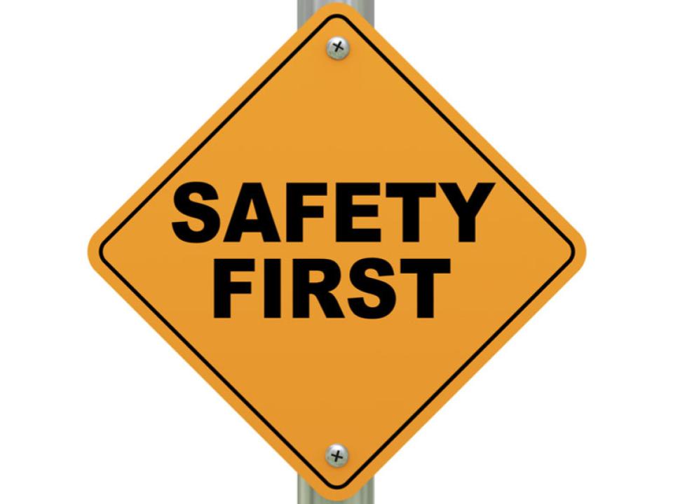
Before beginning lab activities, you must review and understand our safety procedures. This lab follows Biosafety Level 1 (BSL-1) guidelines and includes the use of equipment and materials that must be handled with care. You’ll learn proper lab conduct, emergency protocols (like R.A.C.E. and P.A.S.S. for fire safety), and how to use safety gear such as eye washes, fire blankets, and lab PPE.
All students must follow dress code policies and understand appropriate waste disposal procedures for biohazards, sharps, and general waste. Your safety and the safety of those around you depend on these protocols being followed strictly. Please review all safety materials and confirm your understanding before participating in any lab activities.
Upon completion of this module, you will be able to:- Identify the characteristics and appropriate uses of a Biosafety Level 1 (BSL-1) laboratory, including the types of agents handled and safety precautions required
- Demonstrate proper use and location of essential laboratory safety equipment, including fire extinguishers, fire blankets, safety showers, and eye wash stations
- Describe appropriate personal protective equipment (PPE) and dress code requirements necessary for safe conduct in a BSL-1 lab environment
- Apply proper procedures for handling laboratory equipment, reporting incidents, and maintaining a clean and safe workspace
- Differentiate between waste disposal methods for biohazard, sharps, and regular trash based on the material type and associated risk
To achieve these objectives:
- Read the Introduction & Lab Safety chapter in the lab manual.
- Review the Introduction & Lab Safety 1 PowerPoint slides.
- Complete the Introductions & Lab Safety Quiz before the DUE DATE (see the Course Calendar).
-
Students mustMark as done
Guidelines for how to import content into Moodle
-
Introduction
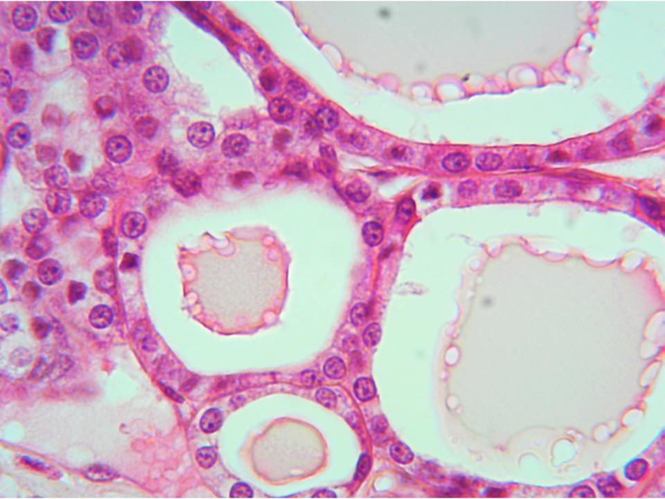
Welcome to the endocrine system lab, the first hands-on module in your Anatomy and Physiology II Laboratory course. During this lab, you will examine the body’s major endocrine glands—such as the pituitary, thyroid, adrenal glands, pancreas, and gonads—and learn about the hormones they produce. Using anatomical models, microscope slides, and interactive activities, you will identify endocrine tissues, observe their microscopic features, and analyze their physiological roles. You’ll also investigate how the endocrine system interacts with other body systems to adapt to internal changes (like stress or blood sugar fluctuations) and external stimuli (such as temperature or environment). This lab will help you draw connections between the structure and function of endocrine organs and the effects of hormonal imbalances. Understanding the endocrine system is essential for recognizing how disruptions in hormone levels can lead to conditions such as diabetes, thyroid disorders, or adrenal insufficiency.
Upon completion of this module, you will be able to:- Describe the overall function of the endocrine system
- Describe the functions of the individual organs and structures of the endocrine system
- Identify the structures of the endocrine system and associated parts on a model, in a picture or diagram, or in a specimen.
- Identify and describe the histologic structures of the endocrine system in a microscope slide.
- List the hormones secreted by the endocrine organs, and identify their site of origin
To achieve these objectives:
- Read Chapter 1 in the textbook and Chapter 1 in the lab manual.
- Complete the Chapter 1 Pre-Lab exercises and Module 1 Quiz before the DUE DATE (see the Course Calendar).
- Complete the Chapter 1 Lab Exercises [in-class or online].
Note the check boxes to the right that help you track your progress: some are automatic, and some are manual. [Delete in subsequent modules.]
Module Pressbooks Resources and Activities
You will find the following resources and activities in this module at the Pressbooks website. Click on the links below to access or complete each item.
-
-
Opened: Wednesday, June 25, 2025, 12:00 AMDue: Wednesday, July 2, 2025, 12:00 AM
-
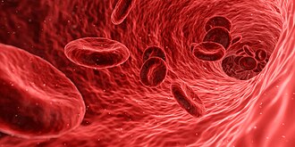
Blood is the body's superhighway, transporting red blood cells, white blood cells, platelets, nutrients, hormones, gases, and wastes. In this module, you will be learning how to identify various components of blood as well as learning about blood typing and why it matters for blood transfusions. You may find yourself using this information in many healthcare fields, including primary care, emergency medicine, laboratories, and blood banking. (Image: Red Blood Cell.jpg, Arek Socha, Pixabay, Creative Commons.)
Upon completion of this module, you will be able to:Describe the functions of blood (Course Objective #3)
Describe the cellular and extracellular components of blood, including plasma and the formed elements (Course Objective #3)
Identify the formed elements of blood in a blood smear (Course Objectives #2 & #3)
Identify blood types and explain the physiological basis of blood types (Course Objective #3)
Be able to perform and interpret results from a basic blood typing test (Course Objective #3 & #4)
To achieve these objectives:
- Read Chapter 2 in the textbook and Chapter 2 in the lab manual.
- Complete the Module 2 Pre-Lab exercises and Module 2 Pre-Lab Quiz before the DUE DATE (see the Course Calendar).
- Complete the Module 2 Lab Exercises [in-class or online].
- Complete the Module 2 Post-Lab Quiz,
Module Pressbooks Resources and Activities
You will find the following resources and activities in this module at the Pressbooks website. Click on the links below to access or complete each item.
-
Opened: Friday, October 17, 2025, 12:00 AMDue: Friday, October 24, 2025, 12:00 AM
-
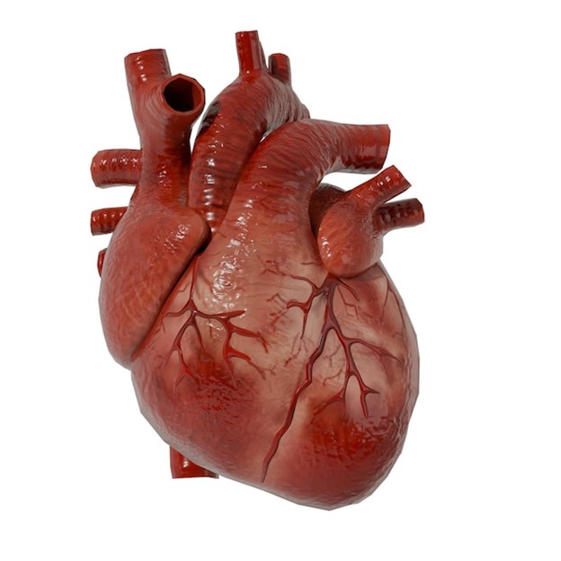
The cardiovascular system is a closed-loop transport system that includes the coordination of the heart and blood vessels. Next to the brain, the heart is considered the most vital organ in the human body. The human heart is a muscular organ with a built-in conducting system located slightly in the center of the thoracic cavity. Anatomical structures of the heart include four chambers (two atria and two ventricles) and contain valves that ensure the blood flows in the correct direction. The pulmonary circuit's primary function is to oxygenate the blood and move blood from the right side of the heart to the lungs and back to the heart. The systemic circuit distributes oxygenated blood with nutrients to body tissues and returns deoxygenated blood to the right side of the heart to repeat the cycle. Blood vessels are a network of conduits for transporting blood. They allow the pulmonary and systemic circuits to work together. Arteries carry blood away from the heart, while veins carry blood to or toward the heart. Capillaries exchange nutrients and gases between the blood and tissues. The network of blood vessels matters because they keep tissues alive by supplying oxygen and nutrients, removing carbon dioxide and waste, supporting the immune system, and regulating body temperature and pH balance.
(Image: https://img.freepik.com/premium-photo/real-human-heart-anatomy-model_1159374-1.jpg.
Upon completion of this module, you will be able to:- Identify and compare anatomical features of the anterior and posterior human heart.
- Describe the internal anatomy of the heart, including the chambers and valves.
- Identify the external anatomy of the heart, anterior and posterior.
- Identify and define cardiovascular system anatomical terms.
- Trace the pathway of blood in the correct sequence.
- Identify the anatomy of cardiac muscle.
To achieve these objectives:
- Read Chapter 3 in the textbook and Chapter 3 in the lab manual.
- Complete the Module 3 Pre-Lab exercises and Module 3 Pre-Lab Quiz before the DUE DATE (View the Course Calendar).
- Complete the Module 3 Lab Exercises.
- Complete the Module 3 Post-Lab Quiz.
Module Pressbooks Resources and Activities
You will find the following resources and activities in this module at the Pressbooks website. Click on the links below to access or complete each item.
-
Opened: Wednesday, December 17, 2025, 11:46 AMClosed: Tuesday, December 30, 2025, 11:46 AM
The pre-lab quiz will acknowledge items you already know about the cardiovascular system.
-
Opened: Wednesday, December 17, 2025, 7:18 AMDue: Tuesday, December 30, 2025, 7:18 AMStudents mustMark as done
-

Figure 4.1. Superficial veins in the upper limb. (Image: OpenStax - Colin Davis)
Introduction
In the cardiovascular system, blood vessels collectively function as a transport network delivering oxygen, nutrients, hormones, and waste products to and from tissues while regulating blood pressure and maintaining homeostasis. Blood vessels have specific functions that allow them to operate as a closed-loop circuit with the heart. Arteries carry blood away from the heart, while veins carry blood to or toward the heart. Capillaries exchange materials between the blood and tissue. The anatomical design of blood vessels allows arteries to function as pressure reservoirs, veins as volume reservoirs, and capillaries as exchange hubs. Knowledge of blood vessel structure, circulation routes, and vessel locations is essential for recognizing how underlying cardiovascular issues may present as external signs or symptoms. This understanding is particularly important in emergency and clinical settings, where rapid identification of internal problems can be critical to patient care. In this laboratory, you will use models, diagrams, and histology slides to study the structures, functions, and locations of major blood vessels.
Upon completion of this module, you will be able to:-
Analyze blood flow through major vessels by determining the vessel from which blood has arrived and the vessel to which it will next travel, including vessels involved in specialized circulatory routes.
-
Identify and explain specialized circulations, including listing the veins of the hepatic portal system and the arteries of the Circle of Willis, and describing the functional significance of each system.
-
Identify and compare blood vessels at the microscopic and macroscopic levels, including recognizing arteries, veins, and capillaries in histology slides; describing the layers of blood vessel walls and their tissues; and correlating structural differences with vessel function.
To achieve these objectives:
- Read the Module 4 Introduction
- Read Lab 4 in the lab manual.
- Take the Module 4 pre-lab quiz.
- Complete the Module 4 lab exercises.
- Take the Module 4 post-lab quiz.
Module Pressbooks Resources and Activities
You will find the following resources and activities in this module at the Pressbooks website. Click on the links below to access or complete each item.
-
-
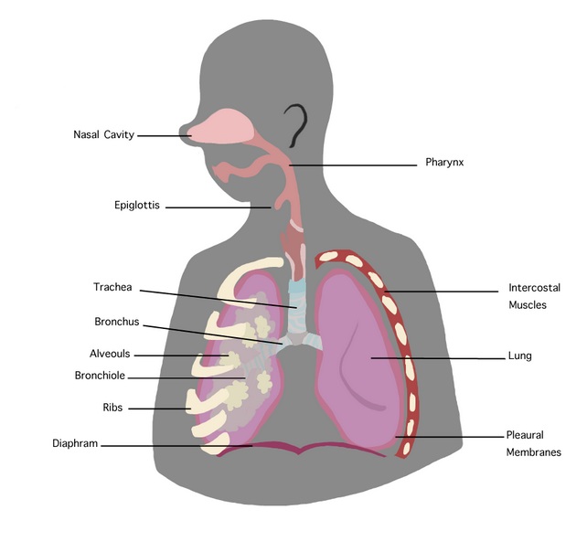
Fig1. Structures of the respiratory system. 2012. Image by Yoshina. CC-BY-3.0
Introduction: The respiratory system regulates pulmonary ventilation to keep blood levels of carbon dioxide, oxygen, and pH within normal limits. In this lab, you'll explore respiratory physiology through two main activities. First, you'll use a simulation to gather data on subjects exposed to hypercapnia (high carbon dioxide) and hypoxemia (low oxygen) to understand how factors like arterial pCO2, pO2, and blood pH affect ventilation. These measurements are essential for diagnosing respiratory, cardiovascular, and urinary conditions. In the second part of the lab, you'll observe and analyze changes in breathing patterns and learn how the respiratory system adjusts to maintain homeostasis.
Upon completion of this module, you will be able to:1. Define the terms pulmonary ventilation, exhalation, inhalation, vital capacity, tidal volume
2. Review the bicarbonate buffering system and the role of the respiratory system in maintaining normal
blood pH3. Define hypercapnia and hypoxemia
4. Discuss the role of arterial pCO2, arterial pO2, and blood pH in regulation of pulmonary ventilation
o How does pulmonary ventilation change in response to changes in blood pH, pCO2, and pO2?
o Why does pulmonary ventilation change in response to changes in blood pH, pCO2, and pO2?
To achieve these objectives:
- Read the Module 5 Introduction
- Complete the Module 5 Pre-Lab exercises and Module 6 Pre-Lab Quiz before the DUE DATE (see the Course Calendar).
- Complete the Module 5 Lab Exercises [in-class or online].
- Complete the Module 5 Post-Lab Quiz
**Please note: the final project should be submitted in this module or the next. Consider this when assigning activities and for grading time.
Module Pressbooks Resources and Activities
You will find the following resources and activities in this module at the Pressbooks website. Click on the links below to access or complete each item.
-
 Image by Rawpixel.com CC0 1.0 Universal.
In this module you will take your midterm exam for this course.
    Read the instructions carefully and take note of any special submission guidelines.
NOTE: Include this module for each exam or major assessment, edited as needed. Include attribution of your image (Image: Title, Author, Source, License).
Upon completion of this module, you will have:
- Read and viewed the [midterm assessment name] instructions
- Scheduled your exam with the proctoring service [if applicable, delete if not needed]
- Prepared for and submitted your midterm assessment [revise as needed]
To achieve these objectives:
- Read and view the contents of "Exam Information and Instructions" [if applicable, delete if not needed]
- Review the [midterm assessment] guidelines in your syllabus to make sure you are ready. Click on [Title of Assessment below] and follow the instructions.
- Log in to the proctoring service and take your exam. [if applicable, delete if not needed]
-
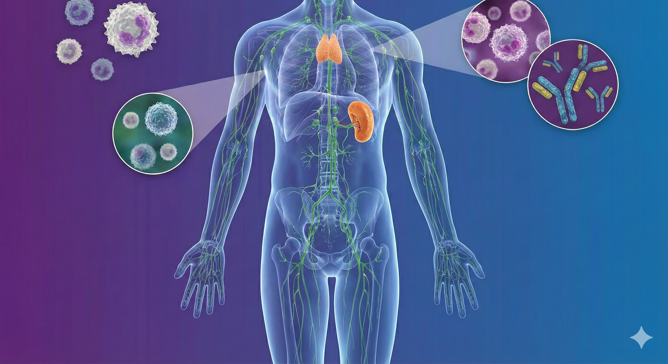
(Image: lymphatic system and immune cells. Image created with Google Gemini).
Introduction: Your Body's Levees and Defense ForceLiving in Louisiana, we know that managing water and protecting our boundaries are full-time jobs. Your body faces similar challenges every day, handled by two closely interconnected networks: the lymphatic and immune systems
. Think of the lymphatic system as your body’s internal drainage commission—much like our pumps and levees, it works to drain excess tissue fluids (interstitial fluid) and return them to the bloodstream to prevent "flooding" or swelling in your tissues . But this system does more than just move fluid; it partners with your immune system to act as a high-tech security team . While the lymphatic vessels transport fluids, the immune system stations specialized cells —such as macrophages, B cells, and T cells—at key checkpoints to filter out pathogens and launch targeted attacks against infections. In this lab, you will explore the anatomy of these defenders. You will trace the "drainage map" of lymph flowing from your feet back to your heart and examine the critical organs where immune cells train and work, such as the thymus, spleen, and lymph nodes . You will also differentiate between your body's immediate "first responders" (Innate Immunity) and its specialized "SWAT team" (Adaptive Immunity) . From understanding how your body handles a rusty nail wound to seeing how vaccines build your defenses against viruses, this lab will help you visualize the life-saving teamwork happening inside you right now . Upon completion of this module, you will be able to:
- Locate and identify major lymph vessels and organs.(C.O. # 3)
- Describe the structure and function of primary and secondary lymphatic organs. (C.O. # 3)
- Discuss the cells of the immune system, how they function, and their relationship with the lymphatic system. (C.O. # 3)
- Identify the specific types of cells that comprise the immune system. (C.O. # 3)
- Differentiate between innate and adaptive immunity. (C.O. # 3)
To achieve these objectives:
- Read the Module 6 Introduction
- Complete the Module 6 Pre-Lab exercises and Module 6 Pre-Lab Quiz before the DUE DATE (see the Course Calendar).
- Complete the Module 6 Lab Exercises [in-class or online].
- Complete the Module 6 Post-Lab Quiz
**Please note: the final project should be submitted in this module or the next. Consider this when assigning activities and for grading time.
Module Pressbooks Resources and Activities
You will find the following resources and activities in this module at the Pressbooks website. Click on the links below to access or complete each item.
- Locate and identify major lymph vessels and organs.(C.O. # 3)
-
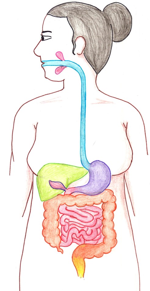
Introduction: The function of the digestive system is to break down the foods you eat, release their nutrients, and absorb those nutrients into the body. Although the small intestine is the workhorse of the system, where the majority of digestion occurs, and where most of the released nutrients are absorbed into the blood or lymph, each of the digestive system organs makes a vital contribution to this process.
In this module, you will be studying the structure of the organs of the digestive system via models, diagrams, histology images, and preserved specimens. You will also review the physiological roles of each structure in accomplishing ingestion, mechanical digestion, chemical digestion, hormone secretion, nutrient absorption, and waste excretion.
Upon completion of this module, you will be able to:
- Describe overall function of the digestive system. (CLO 3)
- Explain the functions of the individual organs and structures of the digestive system. (CLOs 1-4)
- Identify the structures of the digestive system and associated parts on a model, in a picture or diagram, or in a specimen. (CLOs 2-3)
- Identify the histologic structures of the of the digestive system in a microscope slide. (CLOs 2-3)
- Identify the digestive enzymes involved in the digestion of proteins, fats and carbohydrates, and their site of origin. ((CLOs 2-4)
To achieve these objectives:
- Read the Module 7 Introduction
- Read Lab 7 in lab manual.
- Take the Module 7 pre-lab quiz.
- Complete the Module 7 lab exercises.
- Take the Module 7 post-lab quiz.
Module Pressbooks Resources and Activities
You will find the following resources and activities in this module at the Pressbooks website. Click on the links below to access or complete each item.
-
-
-
-
Opened: Monday, July 21, 2025, 12:00 AMDue: Monday, July 28, 2025, 12:00 AMStudents mustMake a submission
-
-
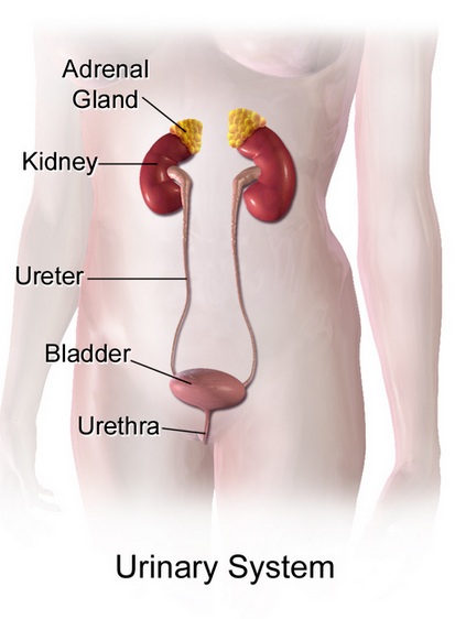
Image credit: A medical illustration depicting urinary system. August 2017. BruceBlus cc-Share Alike-4.0
Welcome to the study of the urinary system, a vital regulatory network that works continuously to maintain the body’s internal balance. Far more than a waste-removal system, the kidneys precisely regulate blood composition by controlling fluid levels, electrolytes, acid–base balance, and blood pressure, while also supporting red blood cell production and Vitamin D activation.
Introduction
The urinary system is essential for preserving the body’s internal stability by constantly monitoring and adjusting the chemical makeup of the blood. Although it is often thought of primarily as the system responsible for producing and excreting urine, its role is much broader. The kidneys regulate water balance, electrolyte levels, acid–base status, and blood pressure, and they also support red blood cell formation and the activation of Vitamin D. Collectively, these functions are fundamental to maintaining homeostasis and ensuring the proper operation of all other organ systems.
These regulatory processes occur at the microscopic level within millions of tiny functional units known as nephrons. Each nephron begins with the glomerulus, a network of capillaries that filters the blood largely according to particle size, preventing cells and most proteins from passing into the filtrate. As the filtrate moves through the nephron’s specialized tubules, specific substances are reabsorbed back into the bloodstream or secreted into the tubule, producing urine with a carefully controlled composition. Despite filtering approximately 200 liters of fluid each day, the kidneys excrete only a small amount, underscoring their exceptional efficiency.
In this laboratory exercise, you will examine both the macroscopic anatomy and the microscopic histology of the urinary system, with a focus on how structure supports function. Through the use of anatomical diagrams and realistic histological images, you will learn to recognize major urinary structures and explore how changes at the cellular level can have widespread effects on the body. As you progress through the lab, keep in mind the guiding question: What happens when this structure fails? This approach will help you see the urinary system not simply as a route for waste elimination, but as a central regulator of the body’s internal environment.
After completing this lab, you should be able to:
- Identify gross and microscopic anatomy of the urinary system.
- Distinguish nephron structures in histological images.
- Relate structure to urine formation and homeostasis.
- Apply anatomical knowledge to clinical scenarios.
To achieve these objectives:
- Read the Modules in the Introduction in Human Anatomy and Physiology II Lecture Moodle course.
- Read and view the materials in Blood and Guts: An OER Approach to Human Anatomy and Physiology II Pressbooks book, linked in yjis module
- Complete the pre-lab, la, and post-lab activities (supplementary videos and lab quiz) included in this laboratory module.
Key Terms:
Urinary System • Kidneys • Nephron • Renal Corpuscle • Glomerulus• Bowman’s Capsule • Filtrate • Proximal ‘Convoluted Tubule (PCT) • Loop of Henle • Distal Convoluted Tubule (DCT)• Collecting Duct • Peritubular Capillaries • Vasa Recta • Filtration • Reabsorption • Secretion • Urine • Erythropoietin (EPO) • Calcitriol • Homeostasis • Renal Cortex • Renal Medulla • Renal Pelvis • Ureter • Urinary Bladder • Urethra • Incontinence
-
Opened: Tuesday, December 16, 2025, 12:00 AMDue: Tuesday, December 23, 2025, 12:00 AM
-
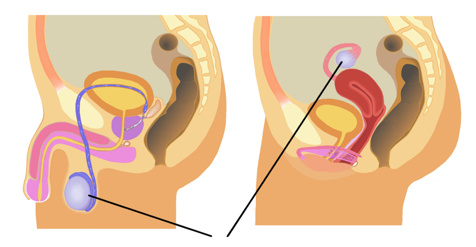
Image: Human Reproductive System, Tsaitgaist and Sciencia58, Wikimedia Commons, CC BY-SA 4.0, https://commons.wikimedia.org/w/index.php?curid=140669671
Welcome to the male and female reproductive system lab module. Here, you will recall content from your lecture materials to assist you in identifying structures (and their functions) of the male and female reproductive systems. You will compare homologous structures between the sexes and identify unique structures to each system. You will review histological samples of reproductive tissues and dissections of these structures in the fetal pig. The identification of structures and functions on 3-D models, in histological samples, and in fetal pigs will provide you with pertinent knowledge you can use in your future career.
Introduction
The male and female reproductive systems complement each other in the creation of a new human. The male reproductive system produces sperm (male gamete), which is required for the fertilization of the ovum (female gamete). Oocytes are produced by the female reproductive system. During intercourse, the male deposits semen into the female's vagina via the process of ejaculation. At this point, the sperm within the semen begin a journey toward the ovum which matured in the ovary and was subsequently released via ovulation. The ovum is collected by the fimbriae and moved into the fallopian tube, where it will ultimately be fertilized by a sperm cell that has traveled through the cervix, uterus, and fallopian tube to reach the ovum. Through fertilization, a zygote will be created and develop into a new human.
Upon completion of this module, you will be able to:
- Identify the internal and external structures associated with the male and female reproductive systems. (CO 3)
- Label primary structures of the male reproductive system. (CO 3)
- Explain the function of each male and female reproductive system structure. (CO 3)
- Recall the process of spermatogenesis. (CO 3)
- Describe the phases of the menstrual cycle and the processes of oogenesis and fertilization. (CO 3)
- Explain lactation and name the structures of the mammary glands. (CO 3)
To achieve these objectives:
- Read the Modules 12-13 Introduction in Human Anatomy and Physiology II Lecture Moodle course.
- Read and view the materials in Modules 12-13 Blood and Guts: An OER Approach to Human Anatomy and Physiology II Pressbooks book, linked in Modules 12 and 13 of the lecture course.
- Complete the pre-lab (8.1-8.5), lab (8.1-8.6), and post-lab activities (supplementary videos and lab quiz) included in this laboratory module.
Overview of Lab Exercises:
- Gross Anatomy: Use models, diagrams, or images to identify the internal and external structures of the male and female reproductive systems.
- Histology: Using provided slides or micrographs (images), identify microscopic structures of the male and female reproductive systems.
- Dissection: Follow the provided protocol to observe the structures of the male and female reproductive systems in the fetal pig.
Module Pressbooks Resources and Activities
You will find the following resources and activities in this module at the Pressbooks website. Click on the links below to access or complete each item.
- Identify the internal and external structures associated with the male and female reproductive systems. (CO 3)
-
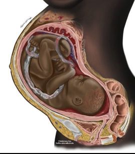
A single fertilized egg cell grows into a fully functional human being during the nine months of pregnancy. In this lab exercise, you will be learning and identifying various structures that develop during embryonic and fetal life. You may use this information in medical fields such as obstetrics, emergency medicine, and family practice (and, of course, if you or a partner become pregnant in the future).
(Image: PregnancyinCrossSection.jpg, Chidiebere Ibe, Creative Commons).
Upon completion of this module, you will be able to:
- Explain early embryonic development and identify the various stages (Course Objective #3)
- Explain fetal development during the three trimesters of gestation (Course Objective #3)
- Explain the anatomy of the placenta and the differences between fetal and postnatal blood circulation (Course Objective #3 & #4)
To achieve these objectives:
- Read Chapter 9 in the textbook and Chapter 9 in the lab manual.
- Complete the Module 9 Pre-Lab exercises and Module 9 Pre-Lab Quiz before the DUE DATE (see the Course Calendar).
- Complete the Module 9 Lab Exercises [in-class or online].
- Complete the Module 9 Post-Lab Quiz,
Module Pressbooks Resources and Activities
You will find the following resources and activities in this module at the Pressbooks website. Click on the links below to access or complete each item.
-

In this module, you will take your final exam for this course. Read the instructions carefully and take note of any special submission guidelines.
Image by Rawpixel.com. CC0 1.0 UniversalUpon completion of this module, you will have:
- Read and viewed the final assessment instructions
- Scheduled your exam with the proctoring service [if applicable]
- Prepared and submitted your final assessment
To achieve these objectives:
- Read and view the contents of the module book "Exam Information and Instructions."
- Review the final assessment guidelines in your syllabus to make
sure you are ready. Click on [Title of Assessment below] and follow the instructions.
- Log in to the proctoring service and take your exam. [if applicable]
Background Colour
Font Face
Font Kerning
Font Size
Image Visibility
Letter Spacing
Line Height
Link Highlight
Text Colour